Join 75,000+ Looksmaxxing Members!

Register a FREE account today to become a member. Once signed in, you'll be able to participate on this site by adding your own topics and posts, as well as connect with other members through your own private inbox.

  • DISCLAIMER: DO NOT ATTEMPT TREATMENT WITHOUT LICENCED MEDICAL CONSULTATION AND SUPERVISION

    This is a public discussion forum. The owners, staff, and users of this website ARE NOT engaged in rendering professional services to the individual reader. DO NOT use the content of this website as an alternative to personal examination and advice from licenced healthcare providers. DO NOT begin, delay, or discontinue treatments and/or exercises without licenced medical supervision. Learn more

Hypothesis UNDERSTANDING FIBROSIS

Askinov

*π’π“π”π…π…Μπ“πŽπ†π€πˆπ*
Joined
Jun 26, 2026
Messages
192
Online time
1d 16h
Reputation
1,976
Location
CHAD LAND

Looksmaxxing Forum Logo

FYBROSIS BY π”„π”–π”Žβ„‘π”‘π”’π”™


So fibrosis comes in two types localized skin fibrosis and medical fibrosis which is caused by skin condition and autoimmune diseases ,

Note : These medical fibrosis are uncontrolled and this requires a medical supervision so asking me about this won't help you .
And now clearing this stuff we can actually move on to the
main topic

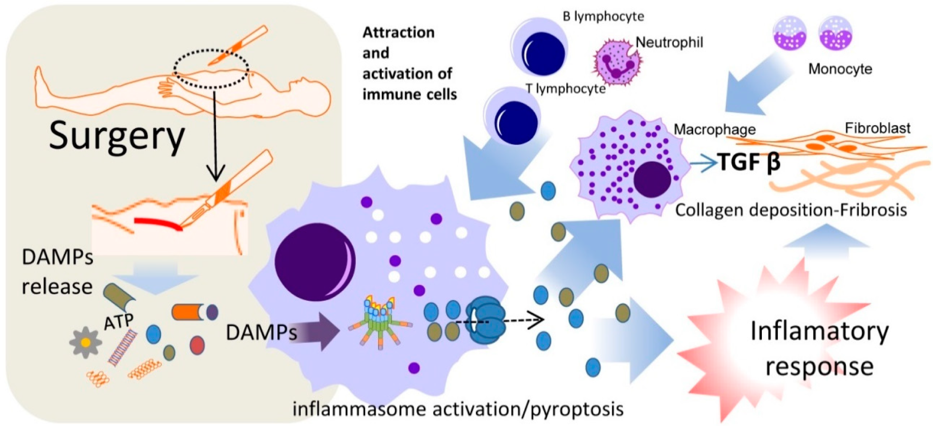
Step 1 ; at 1st the skin experiences a damage any kind of damage it may be major or minor


Damaged epithelial cells (keratinocytes) and endothelial cells (lining local blood vessels) release inflammatory molecules called damage-associated molecular patterns (DAMPs)

Screenshot 2026 08 03 090747


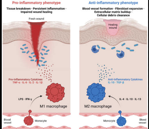
2. Influx of Profibrotic Macrophages

The cellular alarm signals recruit inflammatory white blood cells, mainly M2 macrophages.

In normal healing, M1 macrophages clean the wound and then switch to M2 cells to lay down a repair patch.

In fibrosis, these M2 macrophages stay locked in place, continuously secreting massive amounts of cytokines (cell-signaling proteins).

Screenshot 2026 08 03 085902


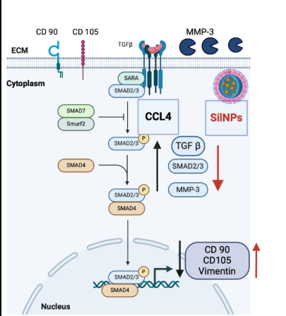
3. TGF-Ξ² Hyper-Activation Loop

The most critical driver of fibrosis is a master-regulator protein called Transforming Growth Factor-Beta 1 (TGF-Ξ²1).

M2 macrophages and damaged cells flood the area with latent TGF-Ξ²1.

Mechanical tension in the stiffening tissue activates this latent TGF-Ξ²1.


Once active, TGF-Ξ²1 binds to receptor proteins on the surface of local resting cells, initiating an intense internal signaling cascade (primarily through the Smad2/3 pathway).
Screenshot 2026 08 03 090513


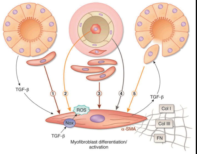
Step4 ; Phenotypic Phenotype Switch: Myofibroblast Differentiation

The TGF-Ξ²1 signaling forces local fibroblasts to undergo a dramatic genetic change. They differentiate into myofibroblasts.

Myofibroblasts express a protein called Alpha-Smooth Muscle Actin (Ξ±-SMA).

Ξ±-SMA gives cells internal muscle-like cables, allowing them to physically grip the surrounding tissue and pull it tightly together.

This cell contraction creates a cycle: more tissue tension β†’ more TGF-Ξ²1 activation β†’ more myofibroblasts.


1785774296291


Step5 ; Extracellular Matrix (ECM) Synthesis and Cross-Linking

The hyper-activated myofibroblasts begin synthesizing structural materials at a rate the body cannot keep up with, flooding the Extracellular Matrix (ECM).

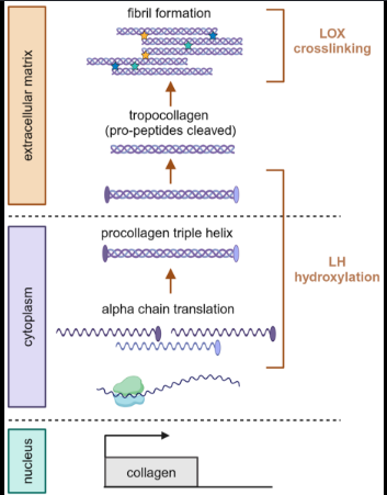
Collagen Shift: Production of Collagen Type III drops which are replaced by massive quantitiy of rigid and dense Collagen Type I.

Cross-Linking: Cells secrete an enzyme called Lysyl Oxidase (LOX). LOX chemically cross-linking the parallel
collagen fibers tightly that the body's natural recycling enzymes MMPs can no longer break them down.

1785774925504


6. Structural Rigidity and Loss of Function

As the densely cross-linked Collagen Type I crowd out the normal cellular space, the microenvironment becomes choked. Blood capillaries are compressed, leading to localized hypoxia (low oxygen), which triggers even more TGF-Ξ²1 release.

The normal cellular geometry is permanently replaced by a rigid, inelastic matrix, resulting in the clinically dense, unyielding structure of fibrotic skin.

And now given this with micro trauma you can sort of create a hard tissue formation via hypertrophic fibrosis which happens after repeated micro trauma you need to do micro damage it overtime to create a hypertrophic scar tissue which is hard and give volume


TLDR

Trauma activates DAMP which activates M2 which sits longer at scar tissue and activate cytokines and TGF -Ξ²1 which They differentiate into myofibroblasts which expresses a protein called lpha-Smooth Muscle Actin (Ξ±-SMA) which grip and surrounding tissue which creates a cycle: more tissue tension β†’ more TGF-Ξ²1 activation β†’ more myofibroblasts. Myofibroblasts floods body with ECM and now the work done


APPLICATIONs ?

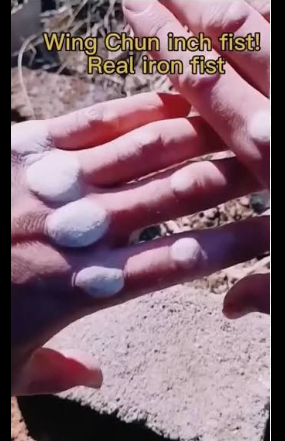
With repeated micro trauma you can create a scar tissue hypertrophic scar tissue which will harden and give volume in the skin

example shaloin monks

1785776408998


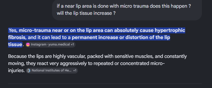
and what i believe is using this method in lip cutting and increasing collagen in lip area so this guy is doing regular cutting which in over time in lip to create a scar tissue which will give the looks of wide lips ( theory )


So to get what i want we need other way of increasing collagen just pure collagen which is discussed in forum

1785776551829


Lip and perioral trauma - PubMed


https://www.sciencedirect.com/topics/medicine-and-dentistry/skin-fibrosis

and final source conor murphy and a nga from org
 
Register to hide this ad

Looksmaxxing Forum Logo

FYBROSIS BY π”„π”–π”Žβ„‘π”‘π”’π”™


So fibrosis comes in two types localized skin fibrosis and medical fibrosis which is caused by skin condition and autoimmune diseases ,

Note : These medical fibrosis are uncontrolled and this requires a medical supervision so asking me about this won't help you .
And now clearing this stuff we can actually move on to the
main topic

Step 1 ; at 1st the skin experiences a damage any kind of damage it may be major or minor


Damaged epithelial cells (keratinocytes) and endothelial cells (lining local blood vessels) release inflammatory molecules called damage-associated molecular patterns (DAMPs)


Screenshot 2026 08 03 090747


2. Influx of Profibrotic Macrophages

The cellular alarm signals recruit inflammatory white blood cells, mainly M2 macrophages.

In normal healing, M1 macrophages clean the wound and then switch to M2 cells to lay down a repair patch.

In fibrosis, these M2 macrophages stay locked in place, continuously secreting massive amounts of cytokines (cell-signaling proteins).


Screenshot 2026 08 03 085902


3. TGF-Ξ² Hyper-Activation Loop

The most critical driver of fibrosis is a master-regulator protein called Transforming Growth Factor-Beta 1 (TGF-Ξ²1).

M2 macrophages and damaged cells flood the area with latent TGF-Ξ²1.

Mechanical tension in the stiffening tissue activates this latent TGF-Ξ²1.


Once active, TGF-Ξ²1 binds to receptor proteins on the surface of local resting cells, initiating an intense internal signaling cascade (primarily through the Smad2/3 pathway).

Screenshot 2026 08 03 090513


Step4 ; Phenotypic Phenotype Switch: Myofibroblast Differentiation

The TGF-Ξ²1 signaling forces local fibroblasts to undergo a dramatic genetic change. They differentiate into myofibroblasts.

Myofibroblasts express a protein called Alpha-Smooth Muscle Actin (Ξ±-SMA).

Ξ±-SMA gives cells internal muscle-like cables, allowing them to physically grip the surrounding tissue and pull it tightly together.

This cell contraction creates a cycle: more tissue tension β†’ more TGF-Ξ²1 activation β†’ more myofibroblasts.


1785774296291


Step5 ; Extracellular Matrix (ECM) Synthesis and Cross-Linking

The hyper-activated myofibroblasts begin synthesizing structural materials at a rate the body cannot keep up with, flooding the Extracellular Matrix (ECM).

Collagen Shift: Production of Collagen Type III drops which are replaced by massive quantitiy of rigid and dense Collagen Type I.

Cross-Linking: Cells secrete an enzyme called Lysyl Oxidase (LOX). LOX chemically cross-linking the parallel
collagen fibers tightly that the body's natural recycling enzymes MMPs can no longer break them down.


1785774925504


6. Structural Rigidity and Loss of Function

As the densely cross-linked Collagen Type I crowd out the normal cellular space, the microenvironment becomes choked. Blood capillaries are compressed, leading to localized hypoxia (low oxygen), which triggers even more TGF-Ξ²1 release.

The normal cellular geometry is permanently replaced by a rigid, inelastic matrix, resulting in the clinically dense, unyielding structure of fibrotic skin.

And now given this with micro trauma you can sort of create a hard tissue formation via hypertrophic fibrosis which happens after repeated micro trauma you need to do micro damage it overtime to create a hypertrophic scar tissue which is hard and give volume


TLDR

Trauma activates DAMP which activates M2 which sits longer at scar tissue and activate cytokines and TGF -Ξ²1 which They differentiate into myofibroblasts which expresses a protein called lpha-Smooth Muscle Actin (Ξ±-SMA) which grip and surrounding tissue which creates a cycle: more tissue tension β†’ more TGF-Ξ²1 activation β†’ more myofibroblasts. Myofibroblasts floods body with ECM and now the work done


APPLICATIONs ?

With repeated micro trauma you can create a scar tissue hypertrophic scar tissue which will harden and give volume in the skin

example shaloin monks


1785776408998


and what i believe is using this method in lip cutting and increasing collagen in lip area so this guy is doing regular cutting which in over time in lip to create a scar tissue which will give the looks of wide lips ( theory )


So to get what i want we need other way of increasing collagen just pure collagen which is discussed in forum


1785776551829


Lip and perioral trauma - PubMed


https://www.sciencedirect.com/topics/medicine-and-dentistry/skin-fibrosis

and final source conor murphy and a nga from org
amazing guide bro, ill read later
 

Looksmaxxing Forum Logo

FYBROSIS BY π”„π”–π”Žβ„‘π”‘π”’π”™


So fibrosis comes in two types localized skin fibrosis and medical fibrosis which is caused by skin condition and autoimmune diseases ,

Note : These medical fibrosis are uncontrolled and this requires a medical supervision so asking me about this won't help you .
And now clearing this stuff we can actually move on to the
main topic

Step 1 ; at 1st the skin experiences a damage any kind of damage it may be major or minor


Damaged epithelial cells (keratinocytes) and endothelial cells (lining local blood vessels) release inflammatory molecules called damage-associated molecular patterns (DAMPs)


Screenshot 2026 08 03 090747


2. Influx of Profibrotic Macrophages

The cellular alarm signals recruit inflammatory white blood cells, mainly M2 macrophages.

In normal healing, M1 macrophages clean the wound and then switch to M2 cells to lay down a repair patch.

In fibrosis, these M2 macrophages stay locked in place, continuously secreting massive amounts of cytokines (cell-signaling proteins).


Screenshot 2026 08 03 085902


3. TGF-Ξ² Hyper-Activation Loop

The most critical driver of fibrosis is a master-regulator protein called Transforming Growth Factor-Beta 1 (TGF-Ξ²1).

M2 macrophages and damaged cells flood the area with latent TGF-Ξ²1.

Mechanical tension in the stiffening tissue activates this latent TGF-Ξ²1.


Once active, TGF-Ξ²1 binds to receptor proteins on the surface of local resting cells, initiating an intense internal signaling cascade (primarily through the Smad2/3 pathway).

Screenshot 2026 08 03 090513


Step4 ; Phenotypic Phenotype Switch: Myofibroblast Differentiation

The TGF-Ξ²1 signaling forces local fibroblasts to undergo a dramatic genetic change. They differentiate into myofibroblasts.

Myofibroblasts express a protein called Alpha-Smooth Muscle Actin (Ξ±-SMA).

Ξ±-SMA gives cells internal muscle-like cables, allowing them to physically grip the surrounding tissue and pull it tightly together.

This cell contraction creates a cycle: more tissue tension β†’ more TGF-Ξ²1 activation β†’ more myofibroblasts.


1785774296291


Step5 ; Extracellular Matrix (ECM) Synthesis and Cross-Linking

The hyper-activated myofibroblasts begin synthesizing structural materials at a rate the body cannot keep up with, flooding the Extracellular Matrix (ECM).

Collagen Shift: Production of Collagen Type III drops which are replaced by massive quantitiy of rigid and dense Collagen Type I.

Cross-Linking: Cells secrete an enzyme called Lysyl Oxidase (LOX). LOX chemically cross-linking the parallel
collagen fibers tightly that the body's natural recycling enzymes MMPs can no longer break them down.


1785774925504


6. Structural Rigidity and Loss of Function

As the densely cross-linked Collagen Type I crowd out the normal cellular space, the microenvironment becomes choked. Blood capillaries are compressed, leading to localized hypoxia (low oxygen), which triggers even more TGF-Ξ²1 release.

The normal cellular geometry is permanently replaced by a rigid, inelastic matrix, resulting in the clinically dense, unyielding structure of fibrotic skin.

And now given this with micro trauma you can sort of create a hard tissue formation via hypertrophic fibrosis which happens after repeated micro trauma you need to do micro damage it overtime to create a hypertrophic scar tissue which is hard and give volume


TLDR

Trauma activates DAMP which activates M2 which sits longer at scar tissue and activate cytokines and TGF -Ξ²1 which They differentiate into myofibroblasts which expresses a protein called lpha-Smooth Muscle Actin (Ξ±-SMA) which grip and surrounding tissue which creates a cycle: more tissue tension β†’ more TGF-Ξ²1 activation β†’ more myofibroblasts. Myofibroblasts floods body with ECM and now the work done


APPLICATIONs ?

With repeated micro trauma you can create a scar tissue hypertrophic scar tissue which will harden and give volume in the skin

example shaloin monks


1785776408998


and what i believe is using this method in lip cutting and increasing collagen in lip area so this guy is doing regular cutting which in over time in lip to create a scar tissue which will give the looks of wide lips ( theory )


So to get what i want we need other way of increasing collagen just pure collagen which is discussed in forum


1785776551829


Lip and perioral trauma - PubMed


https://www.sciencedirect.com/topics/medicine-and-dentistry/skin-fibrosis

and final source conor murphy and a nga from org
First one I actually fully read, does this mean I should cut the corners of my eyes to make them looks better?
 

Users who are viewing this thread

Back
Top